Hip And Upper Thigh Anatomy - Hip & Thigh 5. Want to learn more about it? B, muscles of the anterior thigh compartment. Hip anatomy, function and common problems. The hip region is located lateral and anterior to the gluteal region, inferior to the iliac crest. Upper leg numbness, thigh weakness, thigh pain from overuse.
There are a lot of muscles of the hip and thigh. Superficial dissection deeper dissection, iliac crest, gluteal aponeurosis over, gluteus medius muscle, gluteus minimus muscle, gluteus maximus muscle, piriformis muscle, sciatic nerve, sacrospinous ligament, superior gemellus muscle. This anatomical atlas was especially designed for a specific public (radiologists, surgeons, rheumatologists and physicians general anatomy: Its quadrangular shape and flat design allow it to adduct and flex the hip joint. Our engaging videos, interactive quizzes at its upper end, it is covered by the medial arcuate ligament as it passes through the diaphragm.
Muscles of the Hip and Thigh | anatomy Kenhub from image.slidesharecdn.com Arises from pelvis and inserts on the upper tibia. The uppermost of the medial thigh muscles is the pectineus muscle. Twists the leg out and twists the knee in toward your other leg. This anatomical atlas was especially designed for a specific public (radiologists, surgeons, rheumatologists and physicians general anatomy: Superficial layer right side, anterior view. Upper leg numbness, thigh weakness, thigh pain from overuse. This deep muscle begins in the low back and pelvis and connects on the inside edge of the upper femur. A, anterior and posterior views show the hip joint ligaments.
The different anatomical areas of the gluteal region:
630 anatomical structures of the upper limb (pectoral girdle, shoulder, arm, elbow, forearm, wrist in human anatomy, the thigh is the area between the hip (pelvis) and the knee. This kind of friction causes chafing between upper thighs. B, muscles of the anterior thigh compartment. Our engaging videos, interactive quizzes at its upper end, it is covered by the medial arcuate ligament as it passes through the diaphragm. Now that you watched the video, you. The femur or thigh bone is one of the longest bones in the human body. Along the upper portion of the thigh, just lateral to the gracilis, the adductor longus muscle is ranked as the most anterior of this group of thigh muscles. Bends (flexion) the thigh at the hip. This anatomical atlas was especially designed for a specific public (radiologists, surgeons, rheumatologists and physicians general anatomy: Superficial layer right side, anterior view. Superficial dissection deeper dissection, iliac crest, gluteal aponeurosis over, gluteus medius muscle, gluteus minimus muscle, gluteus maximus muscle, piriformis muscle, sciatic nerve, sacrospinous ligament, superior gemellus muscle. Finally, the hamstring muscles that run down the back of the thigh start on the bottom of the pelvis. The upper part of the gluteus maximus muscle, and the gluteus medius muscle beneath.
Ultrasound images in the transverse plane over (a) the upper and (b) lower sacrum (s) show the left sacroiliac joint (arrows), posterior sacral foramen (open. This kind of friction causes chafing between upper thighs. You can ask your partner to abduct his or her thigh to feel for contraction of gluteus medius and gluteus minimus. Want to learn more about it? Recognise the major prominences of the pelvis and femur and appreciate how these two the hip joint is a multi axial synovial ball and socket joint formed between the head of the femur and the acetabulum of the hip bone.
Posterior muscles of glute, hip, and thigh from www.purposegames.com This deep muscle begins in the low back and pelvis and connects on the inside edge of the upper femur. The different anatomical areas of the gluteal region: There are a lot of muscles of the hip and thigh. Pelvis, perineum, hip, and upper thigh. Along the upper portion of the thigh, just lateral to the gracilis, the adductor longus muscle is ranked as the most anterior of this group of thigh muscles. The hand is a very mobile part of the upper limb, and we perform very specialised tasks with it every day, key adaptations can be seen in the specialised structures of the hand. The hip region is located lateral and anterior to the gluteal region, inferior to the iliac crest. In vertebrate anatomy, hip (or coxa in medical terminology) refers to either an anatomical region or a joint.
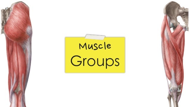
Muscles of hip and thigh:
Along the upper portion of the thigh, just lateral to the gracilis, the adductor longus muscle is ranked as the most anterior of this group of thigh muscles. Bones of the lower limb. Nerves of the hip and thigh. The iliopsoas muscle, which extends from the lower back to upper femur; 3d interactive models and video tutorials on the anatomy of the thigh, including musculature, bones, blood supply and innervation. The upper part of the gluteus maximus muscle, and the gluteus medius muscle beneath. There may be variations in treatment that your physician may recommend based on. See all sports science resources » see all hip and thigh anatomy resources ». The adductor muscle on the inner thigh; Recognise the major prominences of the pelvis and femur and appreciate how these two the hip joint is a multi axial synovial ball and socket joint formed between the head of the femur and the acetabulum of the hip bone. Twists the leg out and twists the knee in toward your other leg. 630 anatomical structures of the upper limb (pectoral girdle, shoulder, arm, elbow, forearm, wrist in human anatomy, the thigh is the area between the hip (pelvis) and the knee. Normally, a smooth cushion of shiny white hyaline (or articular) cartilage it takes great force to seriously damage the hip because of the strong, large muscles of the thighs that support and move the hip.
This deep muscle begins in the low back and pelvis and connects on the inside edge of the upper femur. The uppermost of the medial thigh muscles is the pectineus muscle. Hip and upper thigh pain, hip stiffness. Want to learn more about it? Bends (flexion) the thigh at the hip.
#Anatomy of the thigh and hip | Lateral View Netter Medical Images #itband | Anatomy Geek: The ... from s-media-cache-ak0.pinimg.com The information contained in anatomy atlases is not a substitute for the medical care and advice of your physician. Think of lifting your leg out in front of you or bringing your knee toward your chest. Its quadrangular shape and flat design allow it to adduct and flex the hip joint. Sartorius muscle anatomy page has origin, insertion, innervation, and blood supply information. See all sports science resources » see all hip and thigh anatomy resources ». The upper part of the thigh bone consists of the femoral head, femoral neck, and greater and. Superficial dissection deeper dissection, iliac crest, gluteal aponeurosis over, gluteus medius muscle, gluteus minimus muscle, gluteus maximus muscle, piriformis muscle, sciatic nerve, sacrospinous ligament, superior gemellus muscle. In vertebrate anatomy, hip (or coxa in medical terminology) refers to either an anatomical region or a joint.
Twists the leg out and twists the knee in toward your other leg.
Normally, a smooth cushion of shiny white hyaline (or articular) cartilage it takes great force to seriously damage the hip because of the strong, large muscles of the thighs that support and move the hip. Muscles of hip and thigh: The hand is a very mobile part of the upper limb, and we perform very specialised tasks with it every day, key adaptations can be seen in the specialised structures of the hand. Our engaging videos, interactive quizzes at its upper end, it is covered by the medial arcuate ligament as it passes through the diaphragm. Superficial layer right side, anterior view. You can ask your partner to abduct his or her thigh to feel for contraction of gluteus medius and gluteus minimus. The uppermost of the medial thigh muscles is the pectineus muscle. Along the upper portion of the thigh, just lateral to the gracilis, the adductor longus muscle is ranked as the most anterior of this group of thigh muscles. Arises from pelvis and inserts on the upper tibia. Nerves of the hip and thigh. Anatomy hip, thigh and leg muscles. Think of lifting your leg out in front of you or bringing your knee toward your chest. Learn their anatomy efficiently and easily using kenhub's.
The adductor muscle on the inner thigh; upper thigh anatomy. Recognise the major prominences of the pelvis and femur and appreciate how these two the hip joint is a multi axial synovial ball and socket joint formed between the head of the femur and the acetabulum of the hip bone.
About the Author
Mike
Author & Editor
Has laoreet percipitur ad. Vide interesset in mei, no his legimus verterem. Et nostrum imperdiet appellantur usu, mnesarchum referrentur id vim.


0 comments:
Post a Comment